• Skip to primary navigation
  • Skip to main content
  • Skip to primary sidebar

SATA

Society for the Advancement of Transplant Anesthesia

  • Home
    • For Authors
  • About SATA
    • Committees
      • Abdominal Educational Committee
      • Bylaws Committee
        • SATA Bylaws
      • Cardiothoracic Transplant Committee
        • Cardiothoracic Transplantation Educational Task Force
      • Critical Care Medicine Committee
      • Federation Working Group
      • Fellowship Committee
      • Finance Work Group
      • Membership Committee
      • Newsletter Committee
      • Pediatric Transplant Committee
      • Practice Management Working Group
      • Quality and Standards Committee
      • Research Committee
      • TEE Working Group
      • Vanguard Committee
      • Website Working Group
  • Membership
    • Join Now
  • Education
    • Meetings & Events
      • Meeting
    • Education Materials
      • Article of the Month
      • Protocols / Recommendations / PBLDs
      • Educational Media
      • Transplant Fellowship Information
      • Archive
  • Research
    • SATA Seed Project Funding
    • SATA Publications
  • Newsletter
  • Careers
    • Jobs
    • Post a Job
  • Contact
  • Log In

Case Reports

Implications of the MitraClip procedure for patients requiring LVAD Placement

May 12, 2022 By pacainc

T. Everett Jones, MD, and Yong G. Peng, MD, PhD, FASE, FASA

Department of Anesthesiology, University of Florida College of Medicine, Gainesville, FL


The learning objectives of this PBLD are to:

1) Define the essential evaluation of patient comorbidities before left ventricular assist device (LVAD) placement.

2) Review the impact/implications of a previous MitraClip procedure for patients undergoing LVAD placement.

3) Identify the pertinent transesophageal echocardiography (TEE) findings relevant to LVAD placement.

4) Discuss the strategic approach and management of patients undergoing LVAD placement.


Case Report

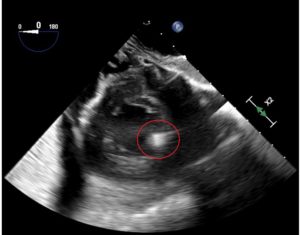
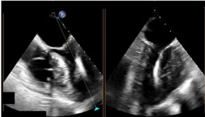
            A 49-year-old man had a past medical history of ischemic cardiomyopathy, coronary artery bypass grafting 10 years prior, chronic renal insufficiency, obesity (BMI 36), congestive heart failure, left ventricular ejection fraction (LVEF) of 20% to 25%, previous St. Jude pacer/automatic implantable cardioverter defibrillator (AICD) for primary prevention, and severe mitral regurgitation after a MitraClip procedure (Abbott, Abbott Park, IL, USA). He presented for placement of a HeartMate III left ventricular assist device (LVAD) as destination therapy. A pre-induction radial arterial line was placed. General endotracheal anesthesia was induced with midazolam, fentanyl, lidocaine, propofol, and rocuronium. The right internal jugular vein was then cannulated using ultrasound guidance with an introducer and a pulmonary artery catheter was floated without incident. His AICD was turned off before the procedure began. Transesophageal echocardiography (TEE) revealed a dilated left ventricle (LV) with global severe hypokinesis and a LVEF of approximately 20%, along with grossly normal right ventricular (RV) function, severe mitral regurgitation, and mild tricuspid regurgitation. A MitraClip was visualized without evidence of mitral stenosis (mean gradient 2 mmHg; Figure 1A and B). A possible residual atrial septal defect (ASD) was identified by color-flow Doppler examination of the interatrial septum. The patient underwent redo sternotomy and was cannulated for cardiopulmonary bypass (CPB). While on CPB, an iatrogenic ASD resulting from his previous MitraClip procedure was closed, the Heartmate III LVAD inflow cannula was placed in the apex of the LV, and the outflow cannula was placed in the ascending aorta. While examining the patient with TEE while he was on CPB, we noted that the inflow cannula was not optimally aligned (Figure 2). The surgeon adjusted the inflow cannula to allow for a more appropriate flow. The LVAD speed was slowly increased in increments of 200 RPMs while the patient was weaned from CPB. A pump speed of approximately 5100 RPMs was eventually reached. The patient was successfully weaned from CPB and protamine was administered. The total CPB time was 147 min. The patient was successfully transitioned to LVAD support without difficulty on infusions of milrinone, epinephrine, norepinephrine, and 40 ppm inhalational nitric oxide (NO). Inflow cannula peak velocities measured between 80 and 100 cm/s on TEE. The patient was transported to the cardiac intensive care unit uneventfully. He was extubated on postoperative day 2.


Discussion

1. Why is preoperative evaluation of patient comorbidities important for LVAD placement?

Preoperative evaluation is vital for ventricular assist device placement. Discussions with the surgeon about the urgency of placement (elective vs emergent), planned surgical approach (sternotomy vs thoracotomy), and likelihood of RV assist device placement, as well as any additional planned surgical procedures is necessary. It is important to determine the etiology of the patient’s cardiac and coexisting diseases, paying particular attention to the patient’s current medications, inotropic infusions, pulmonary artery pressure, and RV function. The presence or absence of an AICD and the management plan for the AICD should be noted. Additionally, take note of the hematocrit; many patients have received erythropoietin stimulating agents preoperatively and removal of autologous blood may be considered.


2. What pertinent preprocedural TEE findings are relevant for LVAD placement?

A systematic TEE examination is essential before LVAD placement, paying particular attention to the assessment of structural and functional abnormalities. Significantly reduced LV contractile function in this population increases the risk of LV thrombus. The presence of a intracardiac shunt such as a patent foramen ovale (PFO), ASD, or ventricular septal defect (VSD) can lead to hypoxia after LVAD placement secondary to right to left shunting. Structural valvular pathologies such as aortic regurgitation or mitral stenosis will negatively affect the function of the LVAD. Close to normal RV systolic function is essential for the success of LVAD placement.


3. How can a history of a MitraClip procedure affect LVAD placement?

The MitraClip procedure is a structural heart intervention that attempts to address severe mitral regurgitation in patients whose comorbidities prohibit an open mitral valve repair or replacement. The procedure requires a trans-septal puncture through the interatrial septum to deliver the mitral clip. The intervention is successful when a significant reduction of mitral regurgitation is achieved without causing mitral stenosis. However, in certain circumstances, mitral clip application may result in mitral stenosis and/or no significant reduction of mitral regurgitation. The ASD (Figure 3) is typically not closed at the conclusion of the procedure. This iatrogenic intracardiac shunt typically closes spontaneously over time, but this is not always the case. If it is present in a patient undergoing LVAD implantation, it is necessary to close the defect.


4. How can TEE guide LVAD inflow and outflow cannula placement?

The preoperative TEE examination should rule out LV thrombus, and intraoperatively it can guide the surgeon to locate an appropriate area to place the inflow cannula at the LV apex. The goal is to directly align the inflow cannula with the mitral valve inflow. The LVAD inflow cannula should avoid angulation toward either the septal wall or the lateral wall. The LVAD inflow cannula velocity should be laminar and approximately 100 cm/s. The LVAD outflow cannula is located in the proximal ascending aorta. TEE assessment should be free of aneurysm, dissection, or significant atheromatous disease at the site of LVAD outflow cannula placement.


5. Should separation from CPB be accomplished by setting the LVAD to full speed immediately?

LVAD devices require constant hemodynamic and echocardiographic monitoring in the immediate post-implantation period during and after weaning from CPB. LVAD function is preload dependent and afterload sensitive. After separation from CPB, ensuring adequate fluid replacement and making slow adjustments to the LVAD speed are strongly recommended. Failure to maintain normovolemia and adequate LV filling can lead to “suction events” (Figure 4), which can lead to acute right heart failure due to changes in the RV geometry. The medical professional should be aware of the potential for suction events and rely on institutional/peer-reviewed protocols to prevent such devastating yet avoidable events.


6. Why is monitoring RV systolic function essential during placement of a LVAD?

Many patients undergoing LVAD placement present with pulmonary arterial hypertension with tenuous RV systolic function. It is essential to constantly monitor RV systolic function during the course of LVAD placement. Particular attention should be paid to avoiding increases in pulmonary vascular resistance. Thus, it is necessary to avoid hypoxia, hypercarbia, significant sympathetic stimulation, hypothermia, and acidosis; these factors may result in an acute increase in pulmonary vascular resistance. Administration of inotropic agents such as milrinone and dobutamine, as well as pulmonary vasodilators such as inhaled nitric oxide or epoprostenol can be helpful to ensure that the RV systolic performance can provide the LV filling necessary to keep up with the increased LV output from LVAD function.


7. What is the pulsatility index?

The pulsatility index (PI) is a measure of the pulse flow through the LVAD device from native heart function. LV contraction leads to an increase in ventricular pressure that increases pump flow; the flow pulses generated are measured and averaged over 15 s. The PI increases with enhancement of LV ejection. The PI can abruptly drop due to a sudden decrease of LV output resulting from arrhythmias or LV preload. Barring a decrease in circulating volume, acute RV failure can also lead to a drop in the PI. 


8. How should fluid resuscitation and vasoactive support be balanced immediately after LVAD implantation?

TEE is an invaluable modality to guide fluid management after LVAD placement.  Appropriate LVAD function demands primary fluid replacement instead of continued increases in vasoactive medications. Echocardiography can not only assess the appropriate alignment of the LVAD inflow cannula at the LV apex, but can also be used to ensure adequate filling of the LV. Hypovolemia and acute RV failure both lead to inadequate LV filling that can result in “suction events.” These events can precipitate acute right heart failure or worsen preexisting right heart failure. TEE is useful to evaluate RV function after LVAD implantation, and if RV function is adequate, then volume replacement should be considered. LVAD devices are afterload sensitive and preload dependent. Therefore, vasoconstrictors should not be the primary choice to rescue hypotension; rather, consideration of volume replacement is warranted. Inotropic agents such as epinephrine, milrinone, or dobutamine should be administered to support RV systolic function to promote LV filling. This strategy will allow for appropriate performance of the LVAD device.


References

  1. Silverton NA, Patel R, Zimmerman J, et al. Intraoperative transesophageal echocardiography and right ventricular failure after left ventricular assist device implantation. J Cardiothorac Vasc Anesth. 2018;32;2096–2103.
  2. Gudejko MD, Gebhardt BR, Zahedi F, et al. Intraoperative hemodynamic and echocardiographic measurements associated with severe right ventricular failure after left ventricular assist device implantation. Anesth Analg. 2019;128;1:25–32.
  3. Baxter RD, Tecson KM, Still S, et al. Predictors and impact of right heart failure severity following left ventricular assist device implantation. J Thorac Dis. 2019;11;6:S864–S870.
  4. Rich JD, Burkhoff D. HVAD flow waveform morphologies: theoretical foundation and implications for clinical practice. ASAIO J. 2017;63:526–535.
  5. Katz WE, Conrad Smith AJ, Crock FW, Cavalcante JL. Echocardiographic evaluation and guidance for MitraClip procedure. Cardiovasc Diagn Ther. 2017;7:616–632.


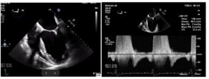
Figure 1. (A) Mid-esophageal four-chamber view reveals the mitral clip between the anterior and posterior mitral valve. (B) Pulsed-wave Doppler reveals a mitral inflow pattern with a peak gradient of 7 mm Hg and mean gradient of 2 mm Hg.


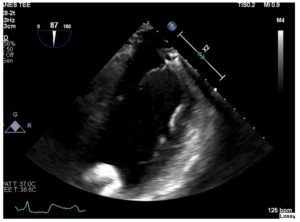
Figure 2. Mid-esophageal two-chamber view reveals malalignment of the LVAD inflow cannula.


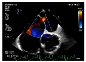
Figure 3. Modified mid-esophageal aortic short-axis view reveals classic example of residual ASD after the MitraClip procedure.


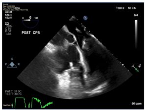
Figure 4. Mid-esophageal four-chamber view demonstrates severe hypovolemia with an obliterated left ventricle.

Filed Under: Case Reports, Education

Retained Cardiac Implantable Electronic Device Material During Orthotopic Heart Transplantation

May 12, 2022 By pacainc

Michael Curtis, MD

University of California, San Francisco

Introduction

            Patients with end-stage heart failure that are scheduled for orthotopic heart transplantation (OHT) frequently present with cardiac implantable electronic devices (CIEDs), such as automatic implantable cardioverter-defibrillators (AICDs) and pacemakers, with or without a cardiac resynchronization therapy function (CRT). AICDs may prevent sudden cardiac death from ventricular arrhythmias that these patients are susceptible to, and pacemakers protect against bradyarrhythmias. CRT may provide both improved cardiac function and symptomatic relief. Given that these are all treating intrinsic properties to the patient’s native heart, CIEDs are typically removed during the intraoperative course of an OHT. Unfortunately, this is not always successfully accomplished – either purposefully or inadvertently – placing the patient at risk of adverse sequelae in the post-operative period.


Incidence of Retained CIED Hardware

            Removing a CIED involves extracting the generator itself from its pocket, as well as the leads that most often transverse the subclavian vein to the superior vena cava (SCV) before implanting into portions of the right atrium (RA) and right ventricle (RV). CIEDs with a CRT function will also have an additional lead positioned in the coronary sinus that will need to be removed. Leads that have been in place for many years, especially for those which include defibrillation coils, may have scar tissue firmly grasping on the leads, which can make extraction a challenging process. Specialized equipment and procedures do exist for these difficult situations, but it is rarely feasible to have all of these tools and skillful operators available during urgent procedures such as an OHT.

            Unfortunately, this means that lead fragments are frequently retained after OHT. In fact, multiple studies at a variety of institutions have shown that 16.2-47.5% of patients getting a CIED removed during OHT still had retained material visible on chest radiography post-operatively.(1-4) The large majority of these leads are in the subclavian vein or SVC, but rare cases of migration to RV and even the left ventricle (LV) have been reported.(5, 6) Risk factors for such retained hardware appear to be: age greater than 50 years, the presence of two or more leads, leads that have been in situ greater than 12 months, and the presence of a dual-coil defibrillator lead.(4)

Outcomes from Retained CIED Hardware

            This retained CIED hardware does appear to increase the risk of upper extremity DVT, and thus in theory related cardiovascular complications like pulmonary embolism.(3, 7) These hardware fragments may also increase the patient’s future radiation exposure, given both the artifacts they may cause during imaging plus the physical obstruction they may pose when using bioptomes for cardiac biopsies in the post-operative period.(3) This may seem insignificant, but these patients are already at increased risk for malignancies than the general population, and this additional exposure may serve to further elevate that risk.(8)

Although there is theoretical concern about retained CIED fragments becoming infected and leading to bacteremia, especially in such an immunosuppressed population, thankfully studies have found that this is a relatively rare event.(3, 4, 7) There is worry that retained hardware will preclude the use of future MRI scans, or at the very least place those patients at risk of thermal injury in the MRI scanner.(7) While the number of OHT patients with lead fragments getting MRIs has been small, there do not seem to be any reported adverse events.(3, 4) Finally, it is not hard to imagine negative consequences from intracardiac migration from lead fragments, such as cerebral or pulmonary embolism, hemopericardium from ventricular perforation, or major valvular damage. However, none of these events were seen in the rare, reported incidences of this situation.(5, 6)

Looking for Retained CIED Hardware

            Careful inspection of removed hardware and the surgical field during OHT is the most important and timely method to either ensure that no fragments are left behind or to raise suspicion that challenging to remove segments may have been retained. Intraoperative transesophageal echocardiography (TEE) may also be able to detect intracardiac fragments or those in the proximal SVC, though without a high index of suspicion, these fragments may not be immediately obvious (Figure 1 and Figure 2) when focused on evaluating donor heart function and anastomotic sites while coming off cardiopulmonary bypass (CPB) with tenuous hemodynamics. Even if immediately detected, it understandably might only prompt a discussion with the surgical team about weighing the relative risk of prolonging CPB time versus planning for later removal endovascularly.

However, as discussed above, the majority of these retained CIED leads remain in the SVC or subclavian veins, which cannot be seen on TEE. Thus, evaluating for their presence on post-operative chest radiography still needs to be an important modality to use during the recovery process.

Conclusion

            Patients presenting for OHT frequently also need concurrent removal of existing CIED hardware. Due to various factors, retention of hardware fragments, especially dual-coil defibrillator leads, is relatively common and can be associated with negative sequelae such as DVTs or, more rarely, infection if not removed. Intraoperative TEE can play an important role in early identification of intracardiac or proximal SVC hardware, but has limitations in evaluating the locations where retention is most frequent. Fragments can be more extensively visualized on other post-operative imaging modalities such as chest radiography, which should be performed in an expedited manner once the patient is stable. No clear guidelines exist for the timing of removing the leads if found. However, many advocate for them to be removed as soon as possible, typically via endovascular approach, in order to eliminate the ongoing risk of complications.(2)

Figure 1 – Transgastric mid-papillary short axis view on transesophageal echocardiography (TEE) after orthotopic heart transplant (OHT) demonstrating an echodense object at the location of the anterolateral papillary muscle (circled in red above). Additional post-operative imaging clarified that this was a retained cardiac implantable electronic device (CIED) lead fragment.

Figure 2 – Concurrent mid-esophageal four chamber and two chamber views on transesophageal echocardiography (TEE) after orthotopic heart transplant (OHT) showing a linear, echodense object extending from the mitral valve to the anterior left ventricular wall (red arrows above). Additional post-operative imaging clarified that this was a retained cardiac implantable electronic device (CIED) lead fragment.

References

  1. Kim J, Hwang J, Choi JH, Choi HI, Kim MS, Jung SH, et al. Frequency and clinical impact of retained implantable cardioverter defibrillator lead materials in heart transplant recipients. PLoS One. 2017;12(5):e0176925. Epub 20170502. doi: 10.1371/journal.pone.0176925. PubMed PMID: 28464008; PubMed Central PMCID: PMC5413001.
  2. Kusmierski K, Przybylski A, Oreziak A, Sobieszczanska-Malek M, Kolsut P, Rozanski J. Post heart transplant extraction of the abandoned fragments of pacing and defibrillation leads: proposed management algorithm. Kardiol Pol. 2013;71(2):159-63. doi: 10.5603/KP.2013.0009. PubMed PMID: 23575709.
  3. Alvarez PA, Sperry BW, Perez AL, Varian K, Raymond T, Tong M, et al. Burden and consequences of retained cardiovascular implantable electronic device lead fragments after heart transplantation. Am J Transplant. 2018;18(12):3021-8. Epub 20180424. doi: 10.1111/ajt.14755. PubMed PMID: 29607624.
  4. Martin A, Voss J, Shannon D, Ruygrok P, Lever N. Frequency and sequelae of retained implanted cardiac device material post heart transplantation. Pacing Clin Electrophysiol. 2014;37(2):242-8. Epub 20140115. doi: 10.1111/pace.12274. PubMed PMID: 24428516.

Filed Under: Case Reports, Education

Lung transplant management in COVID-19 patients

January 1, 2022 By Sergio

Recognizing inadvertent central venous catheter complications and effective management options for patient with COVID-19 infection requiring bilateral lung transplant


Ahmad R. Parniani, MD and Yong G. Peng, MD, PhD, FASE, FASA

Department of Anesthesiology, University of Florida College of Medicine, Gainesville, FL


Summary: We describe the case of a 50-year-old man with COVID-19 who underwent a successful bilateral lung transplant. We also review the anesthetic management strategies for these patients including the role of ECMO and discuss complications associated with central venous catheter placement for lung transplant.


After reviewing the case, readers will be able to:

  • Identify the selection criteria of patients with severe COVID-19 for lung transplant.
  • Recognize the complications associated with central venous catheter placement for lung transplant.
  • Describe the pathophysiology of cardiac tamponade and management strategies.
  • Understand the role of ECMO in the management of patients with COVID-19.
  • Review the anesthetic challenges of patients infected with COVID-19 who are undergoing lung transplant.


Case   

            We describe the case of a 50-year-old man with a past medical history of prostate cancer and prostatectomy, chronic back pain, hypothyroidism, and hypertension. He tested positive for COVID-19 on April 26, 2021 after developing shortness of breath and cough and was sent home from the emergency department for quarantine. However, his condition deteriorated and he was admitted to the hospital 5 days later. He received a combination of remdesivir, dexamethasone, convalescent plasma, and tocilizumab as recommended medical therapy. Unfortunately, on hospital day 20 he was intubated and underwent a percutaneous tracheostomy on hospital day 34. Due to increasing ventilatory requirements, including positive end-expiratory pressure (PEEP) of 24 cm H2O with 100% fraction of inspired oxygen (FiO2), he was placed in the prone position with little improvement. The patient was subsequently placed on venovenous extracorporeal membrane oxygenation (VV-ECMO) on hospital day 60 and transferred to our facility to be evaluated for lung transplant. Given his worsening right ventricular function, he underwent conversion of VV-ECMO to veno-arterio-venous ECMO (VAV-ECMO) on hospital day 64. Epoprostenol and inhaled nitric oxide were continued to treat his pulmonary hypertension and support his worsening right ventricular systolic function. Continuous veno-venous hemofiltration was also initiated given his worsening acute kidney injury.

The patient underwent lung transplant evaluation during his hospital stay and was listed as a lung transplant candidate on August 27, 2021. On September 26, 2021, he underwent a bilateral lung transplant. He was transported to the operating room on VAV-ECMO. Anesthesia was induced with 250 mcg of fentanyl, 130 mg of propofol, and 100 mg of rocuronium. He was intubated orally with a 41-Fr left double-lumen endotracheal tube and the tracheostomy was removed. A transesophageal echocardiography (TEE) probe was inserted for intraoperative monitoring.

Before central venous catheter placement, the left side of his neck was scanned with the ultrasound probe because the ECMO cannulation was in place in his right internal jugular (IJ) vein. However, we could not identify the patent lumen of the left IJ vein (Figure 1).

Figure 1.

As a result, we decided to obtain central venous access through the left subclavian vein. A 9-Fr introducer catheter was successfully placed after one attempt and no resistance was encountered during the insertion. Shortly after the central venous catheter placement, the patient became hemodynamically unstable and unresponsive to multiple vasopressor boluses, including epinephrine. We administered fluids and medication boluses through the newly placed left subclavian central venous catheter. TEE was immediately used to assess his cardiac function and showed an enlarging pericardial effusion with tamponade physiology. At this point, the surgical team was immediately called and resuscitation with intravenous (IV) fluids and epinephrine was continued. We noticed that our boluses of epinephrine were not effective in maintaining his blood pressure, so we switched our infusions and boluses from the central venous catheter into the indwelling peripherally inserted central catheter (PICC). We also started transfusing blood through the ECMO circuit. A peripheral IV was inserted in his left external jugular vein and resuscitation was continued.

A clamshell incision was performed and the surgical exposure entered the pleural cavity on the fourth intercostal space. The pericardium was then opened, and a large amount of blood was evacuated. Afterward, the patient’s condition stabilized, the arterial line tracing became pulsatile once again, and the heart was filling and ejecting appropriately. We noted that the central venous catheter coming from the left subclavian vein tracked outside of the vessel on the thoracic inlet, perforating the pericardium and creating a small tear on the adventitia of the aorta (Figure 2).

Figure 2.

Vasa-vasorum from the aorta adventitia was copiously bleeding, which was controlled with bovie coagulation. The patient became hemodynamically stable and TEE confirmed that his bilateral ventricular function was improving. The surgical team could not access his left femoral vein because ultrasonography revealed thrombosis of the vein.  The case proceeded and a bilateral lung transplant was performed successfully. The patient was transported to the intensive care unit (ICU) postoperatively in a hemodynamically stable condition. He was decannulated from ECMO 1 week after his lung transplant and his tracheostomy was eventually removed. He remained stable on room air and was transferred to rehabilitation facility for further recovery.


What are the current medical therapies available for COVID-19 infection?

Randomized placebo-controlled trials have shown that remdsesivir has enhanced the recovery of hospitalized patients.1,2 Another randomized clinical trial demonstrated that a single dose of bamlanivimab (monoclonal antibody) reduced the viral load in outpatients.2 A large randomized controlled trial showed a mortality benefit with dexamethasone in hospitalized patients requiring oxygen.2 A combination of medical treatments is considered the most effective therapy for patients with COVID-19.

What are the potential treatments for refractory hypoxemia in patients with COVID-19 acute respiratory distress syndrome (ARDS)?

Prone positioning should be considered in patients with PaO2:FiO2 <150 mm Hg during respiration and FiO2 of 0.6 despite an appropriate PEEP. However, prone positioning requires equipment and training that may not be available at some medical centers. Inhaled pulmonary vasodilators such as inhaled nitric oxide can also improve oxygenation in refractory respiratory failure. ECMO can also be considered as an alternative rescue therapy for refractory respiratory failure; however, ECMO is associated with an increased risk of bleeding and requires extensive resources and trained staff.1

What are the ventilatory management strategies in patients with COVID-19 who are intubated?

Autopsies performed on patients with severe COVID-19 have revealed the presence of diffuse alveolar damage, which is a hallmark of ARDS. Essential ventilatory management should focus on avoiding further ventilator-induced lung injury. The main goals are reducing alveolar overdistension, hyperoxia, and cyclical alveolar collapse. Lung-protective ventilation is used by setting the ventilator for tidal volume at 6 mL/kg of predicted body weight. To prevent alveolar overdistention, the plateau pressure should not exceed 30 cm H2O. PEEP prevents alveolar collapse and facilitates the recruitment of unstable lung regions.2

What are the proposed criteria for the selection of patients with severe COVID-19 for lung transplant?

The general criteria are age younger than 65 to 70 years, preference for single organ failure, no malignancy, no substance misuse, and postoperative social support. Other criteria include healthy neurocognitive status, general condition (if the patient is participating in physical therapy while hospitalized), and evidence of irreversible lung damage.1

How long after the onset of ARDS secondary to COVID-19 should lung transplant be considered?

The exact time needed to determine irreversibility is not clear, but the recommendation is to wait approximately 6 weeks after ARDS onset. Ideally, when lung recovery is deemed unlikely after ARDS onset, lung transplant should be considered. Exceptions to these criteria include severe pulmonary complications such as severe pulmonary hypertension with concomitant right ventricular failure, refractory nosocomial pneumonias, or recurrent pneumothoraces that cannot be medically managed with or without ECMO.1

What are some of the concerns regarding ongoing infection at the time of transplant and reinfection of the allograft?

The risk of reinfection should be carefully reviewed as part of the pretransplant evaluation workup. Studies suggest that it is rare to detect replicating virus more than 10 days after infection with SARS-CoV-2 even though the PCR result may remain positive for weeks after infectivity. In cases where the PCR remains positive for extended durations, high-cycle thresholds are seen (Ct >24), but infectivity is usually not evident.1

What are some considerations regarding deep sedation and neuromuscular blockade effects on post-transplant outcomes?

As soon as 2 to 3 days after the start of mechanical ventilation, the diaphragm can lose approximately 50% of its fibers. This can have a significant impact on post-transplant recovery. As a result, weaning of sedation and participation in physical therapy should be highly encouraged.1 In addition, if patients can be weaned from sedation, they can actively participate in discussions regarding the direction of their care and therapeutic goals.

Why are patients with COVID-19 more prone to bleeding during their hospital stay?

ARDS is associated with a significantly increased bleeding risk. In addition, prolonged ECMO can lead to platelet dysfunction in a significant portion of patients. Pleural adhesions and the fragile tissue of these patients increase the risk of intraoperative bleeding.2

What is the approach for placing a subclavian central venous catheter using anatomic landmarks?

Starting 2 cm lateral and 2 cm caudal to the bend of the clavicle, a needle is inserted through the skin at a 30° angle toward the sternal notch. We recommend placing a finger of the nondominant hand in the sternal notch to help find the landmark. Once the needle is under the skin, the needle and syringe are lowered to run parallel to but beneath the clavicle. Access to the vein typically happens just beneath the clavicle, but it may be several centimeters under the skin.3 Medical professionals should be alert to possible complications such as pneumothorax, vascular injury leading to hemothorax, and other inadvertent injury to the adjacent thoracic structures.  

How frequent are complications associated with central venous catheter placement? 

Central venous catheter placement is an invasive procedure that requires planning and an organized approach. There are a number of potential complications associated with central venous catheter placement, most commonly infection, bleeding, pneumothorax, hemothorax, and vascular injury. A prospective randomized trial of patients undergoing subclavian central venous catheter placement at the University of Texas examined the rate of complications. There was a 6% rate of misplacement, 3.7% rate of arterial puncture, 1.5% rate of pneumothorax, and 0.6% rate of mediastinal hematoma.4 Another retrospective chart review was performed for all central venous catheters placed between November 1, 2012, and June 30, 2013, at MedStar Washington Hospital Center (MWHC). In that study, the rate of arterial injury was 1.3% of subclavian central venous catheters, 0.4% of IJ central venous catheters, and 1.4% of femoral central venous catheters.4,5

What is the pathophysiology of pericardial tamponade?

The primary abnormality in cardiac tamponade is impaired diastolic filling of the heart. This is caused by increased intrapericardial pressure that leads to compression of the atria and ventricles. Diastolic filling pressures increase and start to equilibrate with all cardiac chambers. Cardiac filling is reduced, resulting in decreased stroke volume, cardiac output, and systemic blood pressure. Compensatory mechanisms attempt to counteract the decrease in stroke volume by increasing systemic vasoconstriction and tachycardia.6

What are the treatment options and hemodynamic goals during management of patients with pericardial tamponade?

Definitive treatment is emergent drainage and/or relief of the pericardial compression. This can be achieved through pericardiocentesis or surgical decompression. The highlights of hemodynamic management include maintaining contractility and systemic vascular resistance with inotropes and vasopressors. Preload should be maintained with IV fluids and avoiding large tidal volumes of positive pressure ventilation. Additionally, reductions in heart rate should be strongly prohibited to preserve cardiac output because these patients have a fixed and reduced stroke volume.6

What is the role of ECMO in the management of patients with COVID-19?

VV-ECMO is a complicated and labor-intensive tool that is used in severe hypoxemic respiratory failure refractory to conventional mainstays of medical therapy including mechanical ventilation with optimal PEEP, neuromuscular blockade, and prone positioning. VA-ECMO is different from VV-ECMO in that it is typically initiated for patients in cardiac or circulatory failure with or without concomitant respiratory failure. VV-ECMO is commonly considered as a bridge to specific endpoints, such as recovery or lung transplant. Unfortunately, VV-ECMO may also become a bridge to nowhere; a careful assessment of the end goals of therapy is warranted.1,6

What are some of the complications associated with ECMO in patients with COVID-19?

ECMO is associated with thrombotic and hemorrhagic complications. A high proportion of patients with COVID-19 develop life-threatening thrombotic complications. In an autopsy series, most of the patients were diagnosed with deep vein thrombosis (DVT) or pulmonary embolisms. The mechanism of this hypercoagulable state is related to the major systemic inflammatory response along with endothelial dysfunction.7 The combination of a prothrombotic state and long-term use of ECMO cannulas can increase the risk of blood clots. In 80% of patients on ECMO, heparin can be used as a systemic anticoagulant. In patients with COVID-19 or heparin-induced thrombocytopenia (HIT), bivalirudin may be considered as an alternative anticoagulation strategy. Activated clotting times (ACTs) of 160 to 180 s for VV-ECMO and 180 to 220 s for VA-ECMO are necessary to avoid thrombotic complications. Other complications include vascular injury, infections, kidney failure, stroke, and mechanical equipment failure. Closely monitoring ECMO function is essential to the success of this unique bridge therapy.8


References

  1. Bharat A, Machuca TN, Querrey M, Kurihara C, Garza-Castillon R Jr, Kim S, Manerikar A, Pelaez A, Pipkin M, Shahmohammadi A, Rackauskas M, Kg SR, Balakrishnan KR, Jindal A, Schaheen L, Hashimi S, Buddhdev B, Arjuna A, Rosso L, Palleschi A, Lang C, Jaksch P, Budinger GRS, Nosotti M, Hoetzenecker K. Early outcomes after lung transplantation for severe COVID-19: a series of the first consecutive cases from four countries. Lancet Respir Med. 2021 May;9(5):487–497. doi: 10.1016/S2213-2600(21)00077-1. Epub 2021 Mar 31. PMID: 33811829; PMCID: PMC8012035.
  2. Berlin DA, Gulick RM, Martinez FJ. Severe Covid-19. N Engl J Med. 2020 Dec 17;383(25):2451–2460. doi: 10.1056/NEJMcp2009575. Epub 2020 May 15. PMID: 32412710.
  3. Braner DA, Lai S, Eman S, Tegtmeyer K. Videos in clinical medicine. Central venous catheterization–subclavian vein. N Engl J Med. 2007 Dec 13;357(24):e26. doi: 10.1056/NEJMvcm074357. PMID: 18077803.
  4. Mansfield PF, Hohn DC, Fornage BD, Gregurich MA, Ota DM. Complications and failures of subclavian-vein catheterization. N Engl J Med. 1994 Dec 29;331(26):1735–1738. doi: 10.1056/NEJM199412293312602. PMID: 7984193.
  5. Bell J, Goyal M, Long S, Kumar A, Friedrich J, Garfinkel J, Chung S, Fitzgibbons S. Anatomic site-specific complication rates for central venous catheter insertions. J Intensive Care Med. 2020 Sep;35(9):869–874. doi: 10.1177/0885066618795126. Epub 2018 Sep 19. PMID: 30231668.
  6. Gravlee GP. Hensley’s Practical Approach to Cardiothoracic Anesthesia. 6th ed. Wolters Klumer; 2018.
  7. Falcoz PE, Monnier A, Puyraveau M, Perrier S, Ludes PO, Olland A, Mertes PM, Schneider F, Helms J, Meziani F. Extracorporeal membrane oxygenation for critically ill patients with COVID-19-related acute respiratory distress syndrome: worth the effort? Am J Respir Crit Care Med. 2020 Aug 1;202(3):460–463. doi: 10.1164/rccm.202004-1370LE. PMID: 32543208; PMCID: PMC7397791.
  8. Huang J, Firestone S, Moffatt-Bruce S, Tibi P, Shore-Lesserson L. 2021 Clinical Practice Guidelines for Anesthesiologists on Patient Blood Management in Cardiac Surgery. J Cardiothorac Vasc Anesth. 2021 Dec;35(12):3493–3495. doi: 10.1053/j.jvca.2021.09.032. Epub 2021 Sep 24. PMID: 34654633.


Filed Under: Case Reports, Education, Lung Transplant

Extracorporeal membrane oxygenation (ECMO) and peripartum emergency

September 9, 2021 By Sergio

Everett Jones, MD, and Yong G. Peng, MD, PhD, FASE, FASA

Department of Anesthesiology, University of Florida College of Medicine, Gainesville, FL

Case Description

This case describes a 20-year-old woman with a history of asthma (controlled with an inhaler) and illicit drug use who had COVID-19 infection about 1 month before her presentation. She also had a cesarean delivery due to breech position with premature rupture of membranes (at 38 weeks’ gestation) 1 month before presentation. The patient had recently begun to develop shortness of breath over time, which was not responding well to her inhaler. Her family member noted during this time that she was experiencing severe dyspnea, orthopnea, and lower extremity edema. Emergency medical services (EMS) were called after the patient became dyspneic and developed chest pain. While en route to the hospital, she became encephalopathic and her airway was deteriorating. EMS placed a laryngeal mask airway (LMA) after two failed intubation attempts. On arrival to the emergency department, she was pulseless and her rhythm appeared to be pulseless electrical activity (PEA). She had return of spontaneous circulation (ROSC) after successful CPR and intubation. Her urine drug screen was positive for amphetamines, cannabinoids, and opiates. Her troponin levels continued to increase throughout the first 2 days of her admission from 3873 pg/mL to 8303 pg/ml. She received broad spectrum antibiotics for persistent leukocytosis secondary to possible pneumonia. A transthoracic echocardiogram (TTE) revealed new biventricular dysfunction with a left ventricular ejection fraction (LVEF) of 15% to 20%. She was taken to the catheterization laboratory, where she underwent left heart catheterization that did not show coronary artery disease. An intra-aortic balloon pump was placed for circulatory support. She was diagnosed with postpartum/COVID-induced cardiomyopathy and taken directly from the catheterization laboratory to the operating room for central veno-arterial extracorporeal membrane oxygenation (VA ECMO) cannulation. 

This content is for SATA Members only

Login To have access to the Content!

Username:
Password:
Register
Lost your password?

Filed Under: Case Reports, Education

Anesthetic Considerations for Lung Transplant of PAH Group1 Patients

May 2, 2021 By Sergio

Case Presentation

A 16-year-old girl with familial pulmonary arterial hypertension (PAH) presented to our institution for consideration of bilateral lung transplant. She was small for her age because of her chronic medical condition and had been taking pulmonary vasodilators for the past several years…

This content is for SATA Members only

Login To have access to the Content!

Username:
Password:
Register
Lost your password?

Conclusion

Concomitant severe PAH and RV systolic dysfunction present a significant clinical challenge for cardiothoracic anesthesiologists, who should be aware of the potential side effects of various PAH treatment options. A balanced anesthetic technique and appropriate vasoactive selection are essential to the success of lung transplant in this patient population.

Article by: T. Everett Jones, M.D., Yong G. Peng, M.D., Ph.D., FASE, FASA

Filed Under: Case Reports, Education, Lung Transplant Tagged With: lung transplant

Primary Sidebar

Information for Authors

President’s Message

Tetsuro Sakai MD

Tetsuro Sakai MD, PhD, MHA, FASA

SATA President

Read more

Contact Us

Copyright © 2023 Society for the Advancement of Transplant Anesthesia (SATA) - Powered by: Top Shelf Design